Yoga and Anterior Pelvic Tilt

The anterior pelvic tilt is a hot topic in yoga and in manual medicine. Some people are overly concerned about the fact that their pelvis is tilting anteriorly without even understanding what it means. Let’s simplify the issues behind this increasingly common postural variation and learn how it can affect other parts of our body.
A normal pelvic angle is 30°; this means that our posterior superior iliac spines (the dimples above our bottoms) are slightly higher than our anterior superior iliac spines (the bones that stick out at the front of our pelvis). This angle can vary a few degrees either anteriorly or posteriorly due to our genetic makeup and even temporarily due to tight and/or weak muscles. An anterior pelvic angle of 40° is considered excessive and will produce a lower back (lumbar spine) curve that is also extreme. Our lumbar spine should have an anterior curve which is known as a lordosis; however, when this curve is excessive it is known as a hyperlordosis, which is not ideal. Cases of increased pelvic angles and lumbar hyperlordoses are very prevalent in today’s society.
Causes of increased lumbar lordosis include:
- Postural deformity
- Lax muscles, especially the abdominal muscles in combination with tight muscles, especially hip flexors or lumbar extensors
- A heavy abdomen, resulting from excess weight or pregnancy
- Compensatory mechanisms that result from another deformity, such as an increased curve in the thoracic spine (mid-back)
- Hip flexion contracture
- Spondylolisthesis (displacement of the vertebra above with relation to the vertebra below)
- Congenital problems, such as bilateral congenital dislocation of the hip
- Fashion (e.g., wearing high-heeled shoes)
Magee, J. David. Orthopedic Physical Assessment: Fourth Edition. Saunders. Toronto. 2002.
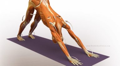
Cause number two from the list above is the reason that affects most. The majority of the population sits for at least eight hours a day while hunched over a desk at work; this can lead to a generic condition known as lower cross syndrome. This disorder consists of the following muscular issues: Weak or inhibited gluteal muscles and abdominals & Tight and shortened hip flexors and lumbar extensors
To better visualize this, observe the illustration that demonstrates lower cross syndrome. Take note of how the two weak/inhibited muscles create one line of the cross and the two tight/shortened muscles create the other line to complete the shape of a cross, hence lower cross syndrome.
When our hip flexors are tight, specifically our psoas, our pelvis rotates forward by the psoas pulling down on the lumbar spine from its attachment sites; this increases our lumbar lordosis and subsequently shortens our lumbar extensor muscles. This is most often seen in combination with weak abdominal and gluteal muscles.
Now that the reasons for this postural condition have been noted, the way to correct it is clear: reverse the causes. However, the distinction between inhibited muscles and weak muscles must be made first before rehabilitation can effectively begin. Inhibited muscles require the re-establishment of correct muscle firing patterns, while weak muscles need to be strengthened. Some individuals have weak muscles that are not inhibited; some have the reverse, and some have both issues to correct. Tight/shortened muscles require lengthening; sometimes stretching is sufficient and sometimes alternative soft tissue treatments such as Active Release Technique® and Graston Technique® are required to decrease scar tissue and increase the range of motion of the particular muscle.
If you are concerned that you may have lower cross syndrome, or simply a pelvic tilt, paying a visit to your manual health care practitioner (sports focused chiropractor, sports physician) will be well worth the time and money. They will be able to diagnose any underlying issues related to this condition and create an appropriate rehabilitation programs specific to the weaknesses and inhibitions they find upon physical examination. They will also be able to reduce scar tissue that may be contributing to your pelvic tilt (anterior or posterior).
Education is the ticket to eliminating these sorts of conditions from society. If we understand how to mitigate the risks for such generic conditions, we will all be much healthier individuals. Here is to learning more about our bodies!
Learn More about Dr. Carla Cupido.
About the Author

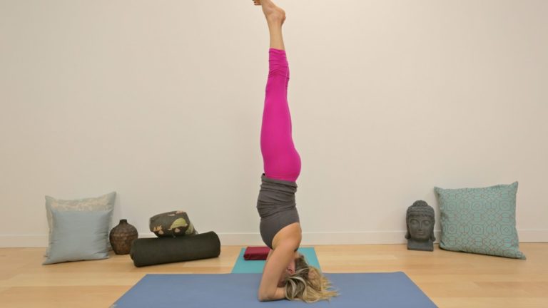
Explore the Anatomy and Correct Alignment of Headstand Pose
Knowledge dissolves fear. With a basic understanding of the structures in your neck, and application of these five keys, one can practice sirsasana safely.
Let’s first take a look at the anatomy, and the neck’s role in our daily life.
The seven little bones of the cervical spine (neck bones) are unique in that they are designed for mobility rather than stability. Like other joints in the body, where stability is sacrificed for mobility, the primary purpose of the C spine in daily life is ease of movement. Therefore, ideal alignment and muscular harmony are particularly important.
The load bearing structures of a cervical vertebrae are the body and two articular facets. A typical cervical vertebral body is approximately two centimeters in diameter depending on the vertebrae (C3 – C7), gender, and individual differences. This is comparable to the diameter of a dime. One may make the comparison of a lumbar vertebral body and cervical vertebral body to the chunky heel of a walking shoe to a high heeled pump. Imagine walking a gravel road in stilettos versus the former.
Another feature worth noting is that the C spine houses the vertebral arteries. Transverse foramen, or holes from top to bottom on the side wings of the bones, house this paired blood vessel which travels up to the brain, taking a rather alarming posterior jog at the top of the neck bones before entering the skull. Symptoms of blocking this small artery include dizziness, blurred vision and occipital headaches. Any lesion compromising the integrity of this passage way is exacerbated by misalignment and the additional and uncustomary weight of your body on the cervical vertebrae in a posture like sirsasana.
Nerves exit the intervertebral foramen (holes in the sides between the neck bones), the branches of which pass laterally between the anterior and middle scalene muscles. These muscles help to hold your head and neck up like guide wires, and provide movement in your neck. Overuse these muscles through misalignment or overload them, and they will become inflamed or tight, possibly pinching the nerves.
How to Safely Practice Headstand (Sirsasana)
Armed with this information, how can you incorporate sirsasana safely into your practice? Headstand or any posture for that matter doesn’t have to look like the pose in your yoga syllabus to start. Practice the actions of the pose in a modification, and you will receive more benefit than forcing the pose.
Here are some important points to practice sirsasana.
1. A strong headstand begins with sensible upright posture.
Carry your upper palate above your physical heart. Assume a natural lordosis in your neck. Your best posture will be your tallest, most easeful posture. Maintain this easeful alignment of your spine in upright yoga postures. If you don’t know what good alignment feels like upright, you won’t know what it feels like upside down.
Practice holding Tadasana in ideal alignment and full attention for several minutes. To simulate the postural muscles further, root down from the outer hips into your feet. Place a block on top of your head while standing, and root up into it from your upper palate as you gently resist. Breathe fully to expand and lengthen your torso. Drop your shoulders away from your ears, and slide the upper arms back to widen the clavicles (collar bones). Invite the ribs back, as this action tends to cause them to splay forward. Breathe into your back, particularly just above the waist.
Practice integrating your body from head to feet with these polar actions of rooting and lifting. When you are in perfect alignment, your body will feel like your favorite pair of walking shoes: No friction, no effort, just ease.
Which brings me to the next key.
2. Stretch your hamstrings and plantar fascia.
To get into any posture, the closer to ideal postural alignment you can get, the less likelihood of injury. To keep your neck safe in headstand, you need to be able to align your entire spine before taking away the support of your feet. In order to achieve this, the back of your legs and soles of your feet must be supple enough to walk into the posture without rounding the lower back and therefore the neck.
3. Apply the rules of progressive overload.
No one walks into a gym and does a clean and jerk with 150 pounds off the bat with no experience. So why would headstand be any different? The neck is accustomed to bearing a mere ten pounds of weight. Add resistance incrementally in weight and duration.
4. Create a stable foundation.
** **When you are ready to do sirsasana, interlace your fingers into prayer hands, with the exception of your pinky fingers. Your pinky fingers should be stacked, overlapping each other front to back. You should be able to see both middle fingers from above but not any of your palm to start–so slightly pronate your forearms. Once you tuck your head into your palms, the tendency is to roll onto the dorsum (back) of your hand. Starting in slight pronation will bring you into neutral alignment once you are in the posture. Now root down through parallel upper arms into the forearms, wrists and hands while keeping the spine neutral and your chest open. Nestle the back of your head into your hands. Distribute the weight between the crown of your head, forearms, wrists and hands.
5. Keep your mouth shut.
This one is mostly for teachers. Although designed primarily to aid in tongue movement and swallowing, the variety of muscles attached to the base of the tongue help to support your neck. Anchor the tongue to the roof of your mouth for additional stability. When it comes to standing on your head, recruit as much help as possible. So teachers, explain your demo first, and don’t speak once you are in the posture.
Precautions and Contraindications
There are precautions and contraindications to performing sirsasana, such as osteoarthritis of the C spine, any autoimmune disease affecting the musculoskeletal system, diabetes, heart condition, degenerated discs, down syndrome, or any other pathology affecting the neck.
However, even with these conditions, one can enjoy many of the benefits of the pose by simply embodying the actions of the pose in a modified form. With patience and keen attention, headstand can be performed safely to benefit your wellbeing.
Naomi Friesen possesses a deep understanding of the physical body through 20 years of teaching movement and anatomy. Students benefit from her knowledge of sound biomechanics by receiving safe and effective instruction. A personal trainer, pilates instructor and lifestyle/weight management coach for 12 years, she now teaches yoga after receiving her yoga instructor certification through Open Source Yoga School. Naomi’s intention is to facilitate connection for herself and students through yoga – connection to Source, connection between the parts of our body, our connection to others.
Website: www.victoriaschoolofyoga.com
Facebook: Victoria School of Yoga